mail_outline sales@mediastorehouse.com
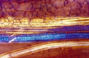
Light Micrograph (LM) of a longitudinal section of stem showing xylem elements of Crown of Thorns (Euphorbia splendens), magnification x1200
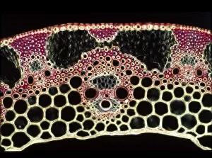
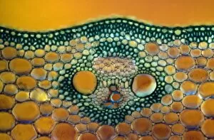
Light Micrograph (LM) of a transverse section of a straw of Wheat showing vascular bundle, cortex and epidermis, magnification x1200
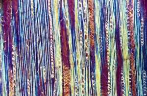
Light Micrograph (LM) of a longitudinal section showing xylem elements of a Ribes sp. stem, magnification x600
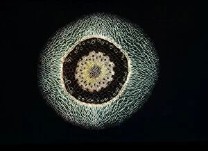
Light Micrograph (LM) of a transverse section of an aerial root of Orchid (Dendrobium sp.), magnification x 30
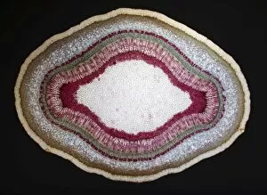
Light Micrograph (LM) of a tranverse section of a stem of a Common (European) Ash tree (Fraxinus excelsior), magnification x12
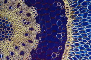
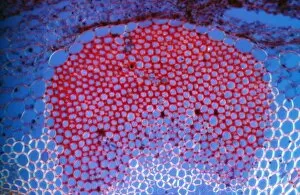
Light Micrograph (LM) of a transverse section of a Maize stem (Zea sp. ) showing vascular bundleLight Micrograph (LM) of a transverse section of a Maize stem (Zea sp.) showing vascular bundle, cortex and epidermis, magnification x600
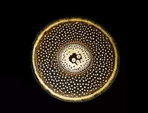
Light Micrograph (LM) of a transverse section of a root of Conifer (Pandanus sp.), magnification x30
Light Micrograph (LM) of a transverse section of an aerial root of Orchid (Dendrobium sp.), magnification x600
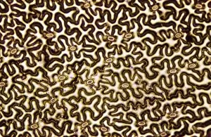
Light Micrograph (LM) of a transverse section showing Sclerenchyma Ground Tissue in Helianthus stem, magnification x 600
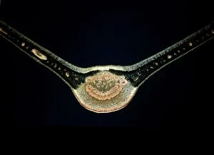
Light Micrograph (LM) of a transverse section of a fig leaf, magnification x 15
Light Micrograph (LM) of a transverse section of a stem of Whisk Fern (Psilotum nudum), magnification x 1200
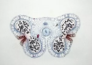
Light Micrograph (LM) of the transverse section of Lilium anthers with mature pollen, magnification x15
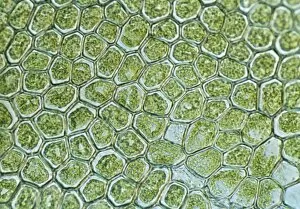
Light Micrograph (LM) of a plant cell chloroplasts, the site where photosynthesis takes place
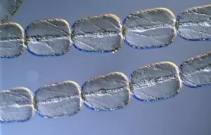
Light Micrograph (LM) of cells from a hair on the stamen of the common spiderwort (Tradescantia)
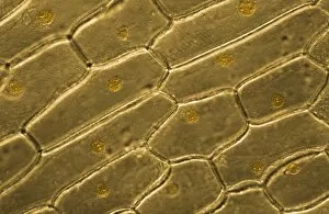
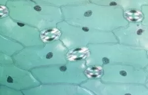
Light Micrograph (LM) of onion skin cells, magnification x 600
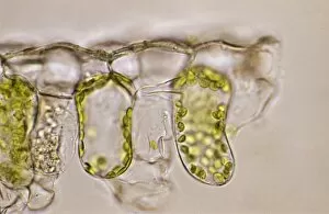
Light Micrograph (LM) of the cellular structure of the non-vascular plant liverwort (Hepatica) showing chloroplasts and oil bodies, magnification x1200
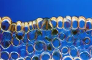
Light Micrograph (LM) of a transverse section of a leaf of a Tulip (Tulipa sp.) showing stomata, magnification x1200
Light Micrograph (LM) of a concentration of cells on the epidermis of a plant showing stomata